Patella Dislocation : Symptoms, Causes, Treatment and Recovery

Prof. Dr. Haluk Çabuk

Prof. Dr. Haluk Çabuk

Professor of Orthopedics & Traumatology
Medically Reviewed Updated November 10, 2026 27 min read

A patella dislocation is a common knee injury that occurs when the kneecap moves out of its normal position in the groove at the end of the thighbone. It often happens after a sudden twist, awkward landing, or direct impact during sports or other activities. The injury can cause sudden knee pain, swelling, and difficulty moving the leg. Some people also experience recurrent dislocations due to underlying knee anatomy or ligament damage.

This article explains what a patella dislocation is and what causes it. You will learn about the different types, common symptoms, how doctors diagnose the injury, and the available treatment options. We also cover when surgery may be necessary, recovery timelines, possible complications, ways to prevent another dislocation, and answers to common patient questions.

Book Appointment

Need a personalized treatment plan?

Book a consultation and receive expert orthopedic guidance.

What Is a Patella Dislocation?

A patella dislocation occurs when the kneecap moves out of its normal position in the groove at the end of the thighbone, called the femoral trochlea. The kneecap usually shifts toward the outer side of the knee, disrupting its normal alignment with the thighbone. It may return to its original position on its own or require a healthcare professional to guide it back.

There are two main forms of patellar displacement:

  • Complete patella dislocation: The kneecap moves entirely out of the trochlear groove, losing its normal alignment with the thighbone.
  • Patellar subluxation: The kneecap partially slips out of its normal position but does not completely leave the groove.

A patella dislocation can happen for the first time after a sudden injury or recur when underlying instability persists. Damage to the ligaments that support the kneecap, particularly the medial patellofemoral ligament (MPFL), can increase the risk of another episode. Anatomical factors, such as a shallow trochlear groove or a high-riding kneecap, can also contribute to patellar instability.
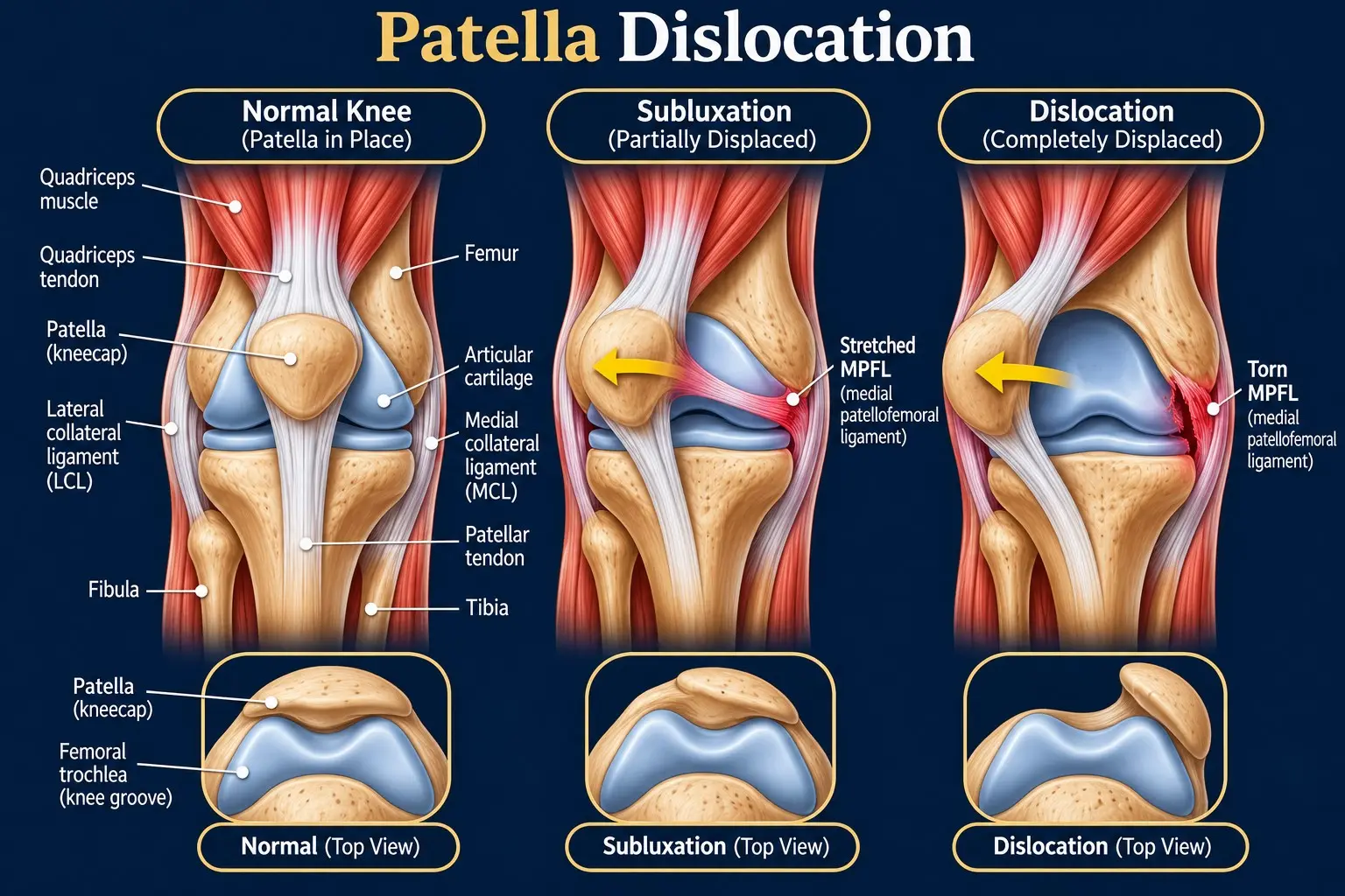
Patella dislocation showing the kneecap displaced from the femoral trochlear groove

How Does the Kneecap Stay in Position?

The kneecap, also called the patella, sits at the front of the knee and moves within a groove at the lower end of the thighbone. This movement helps the quadriceps muscles straighten the knee during walking, running, and other activities. Several bones, ligaments, tendons, and muscles work together to maintain proper kneecap alignment and prevent excessive movement.

The main structures that support patellar stability include:

  • Patella: The kneecap helps protect the front of the knee and improves quadriceps leverage.
  • Femoral Trochlea: This groove at the end of the thighbone guides the kneecap as the knee bends and straightens. A shallow or unusually shaped groove can increase the risk of patellar instability.
  • Medial Patellofemoral Ligament (MPFL): This ligament connects the inner side of the kneecap to the thighbone. It helps prevent the patella from moving too far outward, especially during the early stages of knee bending.
  • Quadriceps Muscles and Tendon: The quadriceps muscles straighten the knee, while their tendon attaches to the upper part of the kneecap. Balanced muscle strength and control help maintain normal patellar tracking.
  • Patellar Tendon: This strong tendon connects the lower part of the kneecap to the shinbone. It transfers force from the quadriceps through the patella to help straighten the leg.
  • Retinacula and Surrounding Soft Tissues: These connective tissues, along with the joint capsule, provide additional support and help control kneecap movement.
  • Hip and Lower-Limb Muscles: The muscles around the hips, thighs, and lower legs help control leg alignment and knee movement during walking, running, jumping, and changes of direction.

These structures work together to maintain patellofemoral stability, which refers to the ability of the kneecap to remain properly aligned as the knee moves. Problems with the trochlear groove, supporting ligaments, muscle control, or overall leg alignment can disrupt normal patellar tracking. This can increase the risk of a dislocated kneecap, particularly during twisting movements or sudden changes in direction.

What Causes a Patella Dislocation?

A patella dislocation can occur when a twisting movement, direct impact, or underlying knee instability forces the kneecap out of its normal position. The injury often involves sudden lateral displacement, meaning the kneecap moves toward the outer side of the knee. Some people experience a dislocation after a significant injury, while others are more vulnerable because of their knee anatomy or previous ligament damage.

What Are the Main Causes of Patella Dislocation?

Several movements and injuries can cause the kneecap to slip out of its normal position.

  • Sudden Twisting Movements: Pivoting sharply or changing direction while the foot remains planted can place excessive stress on the knee. This movement may force the kneecap outward, especially when the supporting structures are weak.
  • Direct Impact: A forceful blow to the kneecap or the inner side of the knee can push the patella out of the trochlear groove.
  • Sports Injuries: Sports such as basketball, soccer, and gymnastics involve sudden stops, jumping, landing, and rapid changes of direction. These movements can increase the risk of patellar dislocation.
  • Falls: Falling directly onto the knee or twisting the leg awkwardly during a fall can displace the kneecap.
  • Awkward Landings: Landing with poor knee control or an unfavorable leg position can place excessive stress on the ligaments and other structures that stabilize the patella.

What Anatomical Factors Can Increase the Risk?

Some people have structural differences that make the kneecap more likely to become unstable. These factors can increase the risk of a first-time dislocation or contribute to repeated episodes.

  • Shallow Trochlear Groove: Trochlear dysplasia occurs when the groove at the end of the thighbone is unusually shallow or poorly shaped. This reduces the bony guidance that helps keep the kneecap aligned.
  • Patella Alta: A high-riding kneecap sits higher than usual and may engage the trochlear groove later during knee bending. This can increase the risk of instability.
  • Abnormal Leg Alignment: Knock-knees, known medically as genu valgum, and certain differences in thighbone or shinbone alignment can affect patellar tracking. An increased Q-angle may also contribute to outward forces on the kneecap.
  • Ligament Laxity: Naturally loose ligaments or generalized joint hypermobility can reduce joint stability. Connective tissue disorders, including Ehlers-Danlos syndrome and Marfan syndrome, may contribute to ligament laxity.
  • Muscle Weakness or Poor Coordination: Weakness or poor control of the quadriceps and hip muscles can affect kneecap tracking. These muscles help control knee movement during everyday activities and sports.
  • Medial Patellofemoral Ligament (MPFL) Injury: The MPFL helps prevent the kneecap from moving too far outward. A tear or stretch injury during a dislocation can leave the knee more vulnerable to another episode.

These causes and risk factors can occur together. A sudden twisting injury may trigger a dislocation in someone with otherwise stable knees, while a person with underlying anatomical differences may experience one during a less forceful movement. Identifying the contributing factors helps doctors choose appropriate treatment and assess the risk of recurrent patellar instability.

What Are the Types of Patella Dislocation?

Patella dislocations can be classified by how far the kneecap moves, the direction of displacement, and whether the injury occurs once or repeatedly. These categories help describe the pattern of patellar instability and identify factors that may affect treatment.

  1. Complete Patella Dislocation: The kneecap moves entirely out of the trochlear groove at the end of the thighbone. It may return to its normal position on its own or require a healthcare professional to reposition it.
  2. Patellar Subluxation: The kneecap partially slips out of its normal groove but retains some contact with the joint. It often returns to its usual position without assistance and may cause a feeling of slipping or instability.
  3. First-Time (Acute) Patella Dislocation: This is the first episode of kneecap dislocation. It often follows a twisting movement, sports injury, fall, or direct impact.
  4. Recurrent Patella Dislocation: The kneecap dislocates repeatedly after previous episodes. Ligament damage, anatomical differences, or persistent patellar instability may contribute to recurrence.
  5. Habitual Patella Dislocation: The kneecap repeatedly moves out of position during a particular movement, such as bending the knee. This pattern may be associated with anatomical abnormalities or muscle imbalances.
  6. Congenital Patella Dislocation: This rare condition involves a kneecap that is displaced from its normal position from birth or early development. It may be associated with underlying developmental or structural differences.
  7. Lateral Patella Dislocation: The kneecap moves toward the outer side of the knee. This is the most common direction of patellar dislocation.
  8. Medial Patella Dislocation: The kneecap moves toward the inner side of the knee. This uncommon pattern may occur after trauma or certain knee surgeries.
  9. Vertical or Rotational Patella Dislocation: The kneecap rotates abnormally or becomes trapped on its edge. This rare pattern usually follows significant trauma and may require specialist treatment to restore its position.

These types describe different features of patellar displacement rather than a single scale of injury severity. A doctor evaluates the displacement pattern, associated ligament or cartilage damage, and any history of previous dislocations to determine the most appropriate treatment.

What Are the Symptoms of a Dislocated Kneecap?

A dislocated kneecap can cause sudden knee pain, swelling, visible deformity, and difficulty moving the leg. Symptoms often begin after a twisting movement, awkward landing, fall, or direct impact. Their severity depends on how far the kneecap moves and whether the injury damages surrounding ligaments, cartilage, or bone.

Common symptoms of a dislocated kneecap include:

  • Sudden, severe knee pain: Intense pain usually develops around the front or outer side of the knee. Moving the leg or putting weight on it may make the pain worse.
  • Visible Kneecap Deformity: The kneecap may appear displaced, sit at an unusual angle, or form a noticeable bump on the outer side of the knee. This sign is more apparent when the patella remains dislocated.
  • Popping Sensation or Sound: You may hear or feel a pop when the kneecap moves out of its normal position. This symptom can occur during the initial injury or as the kneecap returns to place.
  • Swelling and Bruising: The knee may swell rapidly, sometimes within the first few hours. Bleeding or fluid inside the joint can increase swelling, while bruising may develop around the injured area.
  • Difficulty Bending or Straightening the Knee: Pain and swelling can restrict knee movement. The knee may feel stuck or remain in a bent position.
  • Knee Buckling or Instability: The knee may feel loose or unsteady, as though it could give way. Some people also feel that the kneecap might slip out of position again.
  • Difficulty Walking or Bearing Weight: Standing or walking may become painful or impossible because of pain, swelling, or instability. The degree of difficulty varies with the injury and any associated damage.

Symptoms can differ between a complete dislocation and a patellar subluxation, in which the kneecap only partially slips out of its groove. Sometimes, the kneecap returns to its normal position before a medical examination, so visible deformity may no longer be present.

What Should You Do Immediately After a Patella Dislocation?

A patella dislocation can cause sudden pain, swelling, and difficulty moving the knee. You should protect the injured leg and seek urgent medical assessment, even if the kneecap returns to its normal position on its own. The injury may damage surrounding ligaments, cartilage, or bone, even when the kneecap appears normal again.

Follow these steps while arranging medical care.

  1. Stop the Activity Immediately: Stop playing sports, exercising, or walking on the injured leg. Avoid putting weight on the knee until a healthcare professional assesses the injury.
  2. Protect and Support the Knee: Keep the leg supported in its most comfortable position. Avoid unnecessary movement, and do not force the knee to bend or straighten. Do not apply a tight bandage around a visibly displaced kneecap.
  3. Do Not Force the Kneecap Back Into Place: Never push, twist, or pull a displaced kneecap back into position yourself. Incorrect manipulation can worsen damage to the bone, cartilage, ligaments, or nearby structures. A trained healthcare professional should assess and reposition the kneecap safely.
  4. Apply Ice to Reduce Pain and Swelling: Place an ice pack wrapped in a thin towel over the injured knee for 15–20 minutes at a time. Never apply ice directly to the skin. Stop if the skin becomes unusually pale, painful, or numb.
  5. Seek Urgent Medical Assessment: Go to an emergency department if the kneecap remains visibly displaced. You should also seek prompt medical assessment if it returns to position on its own. A doctor may recommend X-rays or other imaging to check for fractures and internal joint damage.
  6. Watch for Emergency Warning Signs: Seek emergency care immediately if you experience any of the following:
    • Severe or rapidly increasing pain or swelling.
    • A kneecap that remains out of position or significant knee deformity.
    • Numbness, tingling, or weakness in the foot.
    • A cold, pale, or discolored foot.
    • An open wound around the injured knee.
    • An inability to actively straighten the knee.

These warning signs may indicate a serious injury that needs immediate treatment. A healthcare professional can assess blood circulation, nerve function, bone alignment, and possible damage to the ligaments or cartilage.

Do not assume the injury is minor just because the kneecap moves back into place. Early assessment helps identify associated injuries and guides the next steps in treatment and recovery.

How Is a Patella Dislocation Diagnosed?

Doctors diagnose a patella dislocation by reviewing how the injury happened, examining the knee, and using appropriate imaging tests. These assessments help confirm the dislocation, evaluate patellar alignment, and identify damage to the surrounding ligaments, cartilage, or bone. The findings also help determine the most suitable treatment.

1. Medical History and Injury Assessment

The doctor asks how the injury occurred and which symptoms developed afterward. This information helps identify the likely cause and determine whether the kneecap has dislocated before.

Your doctor may ask questions such as:

  • Did the injury happen during a twisting movement, fall, jump, or direct impact?
  • Did you see or feel the kneecap move out of position?
  • Did you hear or feel a popping sensation?
  • How quickly did pain and swelling develop?
  • Have you experienced previous patella dislocations or episodes of knee instability?
  • Can you bear weight on the affected leg or straighten your knee?

Your answers help the doctor understand the injury mechanism and assess the possibility of recurrent patellar instability.

2. Physical Examination

The doctor examines the knee for swelling, bruising, tenderness, deformity, and abnormal kneecap positioning. They may check the inner edge of the patella, where the medial patellofemoral ligament (MPFL) can be injured.

The examination may include:

  • Palpation: The doctor gently checks for tenderness around the kneecap and surrounding tissues.
  • Range of Motion: They assess how well you can bend and straighten the knee when it is safe to do so.
  • Patellar Stability: The doctor evaluates kneecap tracking and stability when appropriate.
  • Circulation and Nerve Function: They check blood flow, sensation, and movement in the foot.
  • Extensor Mechanism Assessment: When appropriate, they check whether you can actively straighten the knee.

The doctor avoids forceful movements that could worsen the injury. Some stability tests may be delayed until pain and swelling improve.

3. Knee X-Rays

X-rays are commonly used to assess a suspected acute patella dislocation. They show bone alignment and can reveal fractures or small bone fragments that may have broken away during the injury.

The doctor may request front, side, and specialized kneecap views. These images can also help identify anatomical features that contribute to patellar instability.

Normal X-rays do not rule out cartilage damage or other soft-tissue injuries. Further imaging may be needed if the doctor suspects damage that X-rays cannot show.

4. Magnetic Resonance Imaging (MRI)

An MRI provides detailed images of the ligaments, cartilage, and other soft tissues around the knee. Your doctor may recommend this test if the examination or symptoms suggest associated internal damage.

An MRI can help identify:

  • Tears or damage to the medial patellofemoral ligament (MPFL).
  • Cartilage damage involving the kneecap or thighbone.
  • Osteochondral injuries affecting the cartilage and underlying bone.
  • Loose bone or cartilage fragments inside the knee joint.
  • Other associated soft-tissue injuries.

MRI is particularly useful when significant swelling, persistent pain, or suspected cartilage and bone injuries raise concern. Not every patient needs an MRI, but doctors may recommend one when the findings suggest an associated injury.

5. Additional Imaging When Necessary

A computed tomography (CT) scan may be appropriate when the doctor needs more detailed information about fractures, bone alignment, or rotational abnormalities. It can also help assess anatomical factors in patients with recurrent patella dislocations when the results may influence surgical planning.

The doctor selects imaging tests according to the injury, examination findings, and suspected structural problems. Together, the medical history, physical examination, and imaging results help determine the extent of damage and guide an appropriate treatment plan.

How Is a Patella Dislocation Treated?

Treatment for a patella dislocation aims to restore the kneecap’s position, reduce pain and swelling, and improve knee stability. The approach depends on the severity of the injury, associated damage, and whether the dislocation has happened before. Many first-time dislocations can be treated without surgery when significant cartilage or bone injuries do not require an operation.

1. Restore the Kneecap’s Position

If the kneecap remains dislocated, a trained healthcare professional may perform a procedure called closed reduction. During this procedure, the clinician carefully guides the kneecap back into its normal position. Pain relief or sedation may be provided when necessary.

An untrained person should never attempt to force a visibly displaced kneecap back into place. After reduction, the doctor checks the knee and assesses possible injuries to the surrounding ligaments, cartilage, and bone.

2. Manage Pain and Swelling

Initial treatment focuses on reducing discomfort and protecting the injured knee. Your doctor may recommend several measures to support early recovery.

  • Ice: Apply a cold pack wrapped in a towel for 15–20 minutes at a time to help reduce pain and swelling.
  • Rest: Stop sports and avoid activities that increase pain or place excessive stress on the knee.
  • Elevation: Raise the injured leg when comfortable to help manage swelling.
  • Pain Relief: A healthcare professional may recommend suitable pain medication, including anti-inflammatory medicines when appropriate.

These measures help manage symptoms while the knee begins to recover. Persistent or worsening symptoms may require further assessment.

3. Provide Temporary Knee Support When Needed

A knee brace, splint, or crutches may help protect the knee during early recovery. The doctor determines the type and duration of support based on knee stability, pain, and any associated injuries.

Some patients need only brief support, while others require longer protection. Prolonged immobilization can cause joint stiffness and muscle weakness. Your treatment plan should allow safe knee movement when appropriate.

4. Begin Physiotherapy and Rehabilitation

Physiotherapy helps restore knee movement, rebuild muscle strength, and improve patellar stability. A physiotherapist introduces exercises gradually according to pain, swelling, and healing progress.

A rehabilitation program may include:

  • Range-of-Motion Exercises: These exercises help restore comfortable knee bending and straightening.
  • Quadriceps Strengthening: Strengthening the muscles at the front of the thigh helps improve knee control.
  • Hip and Gluteal Strengthening: These exercises support better leg alignment and movement control.
  • Balance Exercises: Balance training improves coordination and helps develop better knee stability.
  • Functional Training: Exercises prepare you for everyday activities, including walking, climbing stairs, and eventually returning to sports.

Your physiotherapist adjusts the program as strength, mobility, and stability improve. Returning to demanding activities too early may increase the risk of another dislocation.

5. Assess Whether Surgery Is Necessary

Not every patient with a dislocated kneecap needs surgery. Doctors often recommend non-surgical treatment for a first-time dislocation without significant associated damage. Surgery may be appropriate when structural injuries or persistent instability make conservative treatment less suitable.

A surgical assessment may be necessary for:

  • Recurrent Dislocations: The kneecap repeatedly slips out of position despite appropriate treatment.
  • Osteochondral Fractures: Displaced fragments of cartilage and underlying bone may require fixation or removal.
  • Persistent Instability: The kneecap remains unstable despite appropriate rehabilitation.
  • Significant Anatomical Abnormalities: Structural differences contribute to repeated dislocations and may require correction.

The decision depends on imaging findings, symptoms, age, activity level, knee anatomy, and personal goals. Your orthopedic specialist can explain the benefits and risks of each option.

6. Consider Surgery for Significant or Recurrent Instability

When surgery is necessary, the surgeon selects a procedure based on the underlying cause of patellar instability. Common surgical options include:

  • Medial Patellofemoral Ligament (MPFL) Reconstruction: This procedure reconstructs the ligament that helps prevent the kneecap from moving too far outward. Surgeons commonly consider it for recurrent lateral patellar instability and selected cases with significant associated injury.
  • Tibial Tubercle Osteotomy: This procedure repositions the attachment point of the patellar tendon to improve kneecap alignment in selected patients.
  • Trochleoplasty: This procedure reshapes an unusually shallow or malformed groove at the end of the thighbone. Surgeons reserve it for carefully selected patients with significant trochlear dysplasia.

Some patients also need treatment for associated cartilage or bone injuries. The most appropriate procedure depends on the specific injury, knee anatomy, and degree of instability.

The main goal of treatment is to restore stable knee function and reduce the risk of another dislocation. Early medical assessment and an individualized rehabilitation plan help guide recovery, whether treatment involves physiotherapy alone or surgery.

What Complications Can Occur After a Patella Dislocation?

A patella dislocation can damage the cartilage, ligaments, and bone surrounding the kneecap. Some people recover without lasting problems, while others develop persistent pain, reduced knee movement, or recurrent instability. The risk of complications depends on the initial injury, associated damage, and whether further dislocations occur.

Possible complications include:

  • Cartilage Damage: During a dislocation, the kneecap may strike the thighbone and damage the cartilage covering the joint surfaces. This damage can cause persistent pain, swelling, and discomfort during knee movement.
  • Osteochondral Fractures: A dislocation may break a fragment of cartilage and the underlying bone away from the joint surface. Some fragments remain inside the knee and may require surgery, depending on their size, location, and stability.
  • Recurrent Patella Dislocation: The kneecap may slip out of position again, particularly when supporting ligaments are damaged or anatomical factors increase instability. Previous dislocations, a shallow trochlear groove, and a high-riding kneecap can increase this risk.
  • Persistent Patellar Instability: The kneecap may continue to feel loose or unstable, even without another complete dislocation. This can cause apprehension during movement and limit participation in everyday activities or sports.
  • Loose Bodies in the Knee: Detached cartilage or bone fragments may move within the joint. These fragments can cause catching, locking, repeated swelling, or restricted movement. Symptomatic loose bodies may require further assessment and treatment.
  • Persistent Pain and Swelling: Some people experience ongoing discomfort after the kneecap returns to its normal position. Ligament injuries, bone bruising, cartilage damage, or joint inflammation may contribute to these symptoms.
  • Reduced Knee Movement and Muscle Weakness: Pain, swelling, and prolonged immobilization can lead to stiffness and quadriceps weakness. These problems may interfere with walking, climbing stairs, and returning to normal activities.
  • Patellofemoral Arthritis: Cartilage damage or repeated dislocations may increase the risk of arthritis in the joint between the kneecap and thighbone. Symptoms can include pain at the front of the knee, stiffness, and discomfort during squatting or stair climbing.

These complications are possible, but they do not affect everyone who experiences a patella dislocation. Appropriate medical assessment, individualized treatment, and rehabilitation can help restore knee function and address instability.

Seek further medical evaluation if you experience persistent pain, repeated dislocations, or locking of the knee. These symptoms may indicate an associated injury that needs additional treatment.

How Long Does It Take to Recover From a Patella Dislocation?

Recovery from a patella dislocation depends on the severity of the injury, associated damage, treatment, and rehabilitation progress. A first-time dislocation treated without surgery may allow a return to everyday activities within 6–8 weeks. Returning to high-impact sports often takes 3–4 months or longer. Recurrent dislocations, significant cartilage injuries, or surgical treatment can extend recovery to 4–6 months or more.

What Are the Stages of Recovery After a Patella Dislocation?

Recovery usually progresses through several stages. The timing varies according to pain, swelling, knee stability, and healing.

  1. Symptom Management: First Few Days

    The initial goal is to reduce pain and swelling while protecting the injured knee. Treatment may include ice, appropriate pain relief, leg elevation, and temporary knee support. Your doctor may recommend a brace or crutches, depending on the injury and your ability to bear weight safely.

  2. Restoring Mobility: First 1–2 Weeks

    As pain and swelling improve, you may begin gentle knee movements and prescribed rehabilitation exercises. Your healthcare professional will determine when to increase weight-bearing and whether you need continued bracing. The timing depends on knee stability and any associated injuries.

  3. Strengthening and Stability: Following Initial Healing

    Physiotherapy focuses on restoring knee movement and strengthening the quadriceps, hip, and gluteal muscles. Balance and coordination exercises also help improve kneecap control. Your physiotherapist adjusts the program according to your strength, symptoms, and response to exercise.

  4. Gradual Return to Daily Activities and Sports

    Many people with an uncomplicated first-time dislocation return to everyday activities within 6–8 weeks. Running, jumping, pivoting, and other high-impact activities place greater demands on the knee and commonly require 3–4 months or longer.

    You should return to sports only when your knee has regained adequate strength, stability, movement, and functional control. Your healthcare professional can assess your readiness and help reduce the risk of another injury.

How Does Recovery Differ Between Patients?

Recovery timelines vary depending on the injury and the treatment required.

  • First-Time Dislocation Without Surgery: Everyday activities often become manageable within 6–8 weeks, although complete functional recovery may take longer.
  • Recurrent Dislocation or Significant Associated Damage: Persistent instability, ligament injuries, or cartilage and bone damage may extend recovery and require additional treatment.
  • Recovery After Surgery: Rehabilitation commonly takes 4–6 months or longer, depending on the procedure and healing progress. Complex injuries or bone realignment procedures may require up to a year.

A patella dislocation affects the kneecap, while a true knee joint dislocation involves displacement between the thighbone and shinbone. A true knee joint dislocation is a much more serious injury that can damage blood vessels, nerves, and multiple ligaments. Recovery may take 9–15 months or longer, depending on the injuries and treatment required.

These timelines are estimates, not guarantees. Follow your orthopedic specialist's and physiotherapist's guidance throughout recovery. Returning to demanding activities before your knee regains adequate strength and stability can increase the risk of another dislocation.

How Can You Prevent Another Patella Dislocation?

Preventing another patella dislocation involves strengthening the muscles around the knee, improving movement control, and following an appropriate rehabilitation program. The risk of recurrence depends on the initial injury, previous dislocations, ligament damage, and individual knee anatomy. These strategies can help reduce the risk, although they cannot guarantee that another dislocation will never occur.

  • Strengthen the Quadriceps and Hip Muscles: Strong thigh and hip muscles help control kneecap movement and maintain lower-limb alignment. A physiotherapist may recommend exercises such as straight-leg raises, clamshells, wall sits, and step-ups. These exercises help improve muscle strength, coordination, and knee control.
  • Complete Your Rehabilitation Program: Follow your prescribed physiotherapy plan and avoid returning to sports or demanding activities too early. Rehabilitation should gradually restore knee movement, muscle strength, flexibility, balance, and stability. Stopping exercises too soon may leave the knee less prepared for physical activity.
  • Use a Knee Brace and Appropriate Footwear: A patella-stabilizing brace may provide additional support during selected activities when recommended by a healthcare professional. Choose well-fitting footwear that suits your activity and provides appropriate support. Braces and footwear cannot correct every underlying cause of patellar instability.
  • Improve Athletic Technique and Warm Up: Perform a suitable warm-up before sports, including controlled movements that prepare your muscles and joints for activity. During jumping and landing, keep your knees aligned with your feet and avoid uncontrolled inward knee movement. When changing direction, use controlled footwork rather than twisting sharply on a planted leg.
  • Return to Sports Gradually: Increase training intensity as your strength, movement, and confidence improve. A physiotherapist or sports medicine professional can assess your balance, single-leg control, landing technique, and readiness for running, jumping, and pivoting. Do not return to full activity based on time alone.
  • Address Persistent or Recurrent Instability: Consult an orthopedic specialist if your kneecap continues to dislocate despite appropriate rehabilitation. A shallow trochlear groove, high-riding patella, persistent ligament damage, or abnormal leg alignment may contribute to repeated episodes. Selected patients may benefit from procedures such as medial patellofemoral ligament (MPFL) reconstruction, trochleoplasty, or tibial tubercle osteotomy, depending on the underlying cause.

Consistent rehabilitation and appropriate movement training can help improve knee stability and reduce recurrence risk. If you have experienced repeated dislocations, an orthopedic assessment can help identify contributing factors and determine whether additional treatment is needed.

FAQs About Patella Dislocation

1. Can a Dislocated Kneecap Heal on Its Own?

A dislocated kneecap may return to its normal position without assistance, and symptoms can gradually improve. Still, suspected patella dislocation requires medical assessment because associated ligament tears, cartilage damage, or fractures may need treatment. Returning to position on its own does not guarantee that the knee is fully healed.

2. When Can You Return to Sports After a Patella Dislocation?

Returning to sports depends on knee strength, stability, range of motion, and functional recovery. Many patients need three to four months or longer before resuming high-impact sports. Your doctor or physiotherapist should assess your readiness through functional testing and confirm that you can perform sports-specific movements safely.

3. Can You Walk With a Dislocated Kneecap?

Walking may be difficult or impossible when the kneecap remains dislocated because of pain, swelling, and instability. Some people can walk after the kneecap returns to position, but this does not rule out significant damage. Avoid putting weight on the injured leg until a healthcare professional assesses it.

4. How Painful Is a Patella Dislocation?

A patella dislocation often causes sudden, intense pain around the front or outer side of the knee. Swelling, tenderness, and difficulty bending or straightening the leg may follow. Pain levels vary, and the intensity alone cannot determine whether the injury has damaged cartilage, ligaments, or bone.

5. What Is the Difference Between a Patella Dislocation and a Knee Dislocation?

A patella dislocation occurs when the kneecap moves out of its normal groove at the end of the thighbone. A true knee joint dislocation involves displacement between the thighbone and shinbone. A knee joint dislocation is a serious emergency because it can damage major blood vessels, nerves, and ligaments.