
Specialties › Knee & Leg › Patellar Fracture

A patellar fracture is a break in the kneecap, the bone at the front of the knee that protects the joint and helps straighten the leg. It can occur after a direct blow to the knee, a fall, or a sudden, forceful quadriceps contraction. The injury may cause pain, swelling, bruising, and difficulty walking or straightening the knee.
This article explains what patellar fractures are and what causes them. You will learn about their different types, common symptoms, how doctors diagnose them, and the available treatment options. We also cover when surgery may be necessary, how patellar fractures are treated, possible complications, recovery timelines, how to care for your knee during recovery, and answers to common patient questions.
Book a consultation and receive expert orthopedic guidance.
A patellar fracture is a break in the patella, the bone at the front of the knee that helps protect the joint and supports knee extension. It is commonly known as a broken kneecap or fractured kneecap.
The patella connects with the quadriceps muscles and patellar tendon to help straighten the knee. A fracture can cause pain, swelling, and bruising, making walking, bending the knee, or straightening the leg difficult. Severe fractures may disrupt the knee’s extensor mechanism, which can prevent you from actively straightening your knee.
A patellar fracture can range from a small, stable crack to a displaced break in which bone fragments separate. Treatment depends on the fracture pattern, the position of the fragments, and whether the knee’s straightening mechanism remains intact.
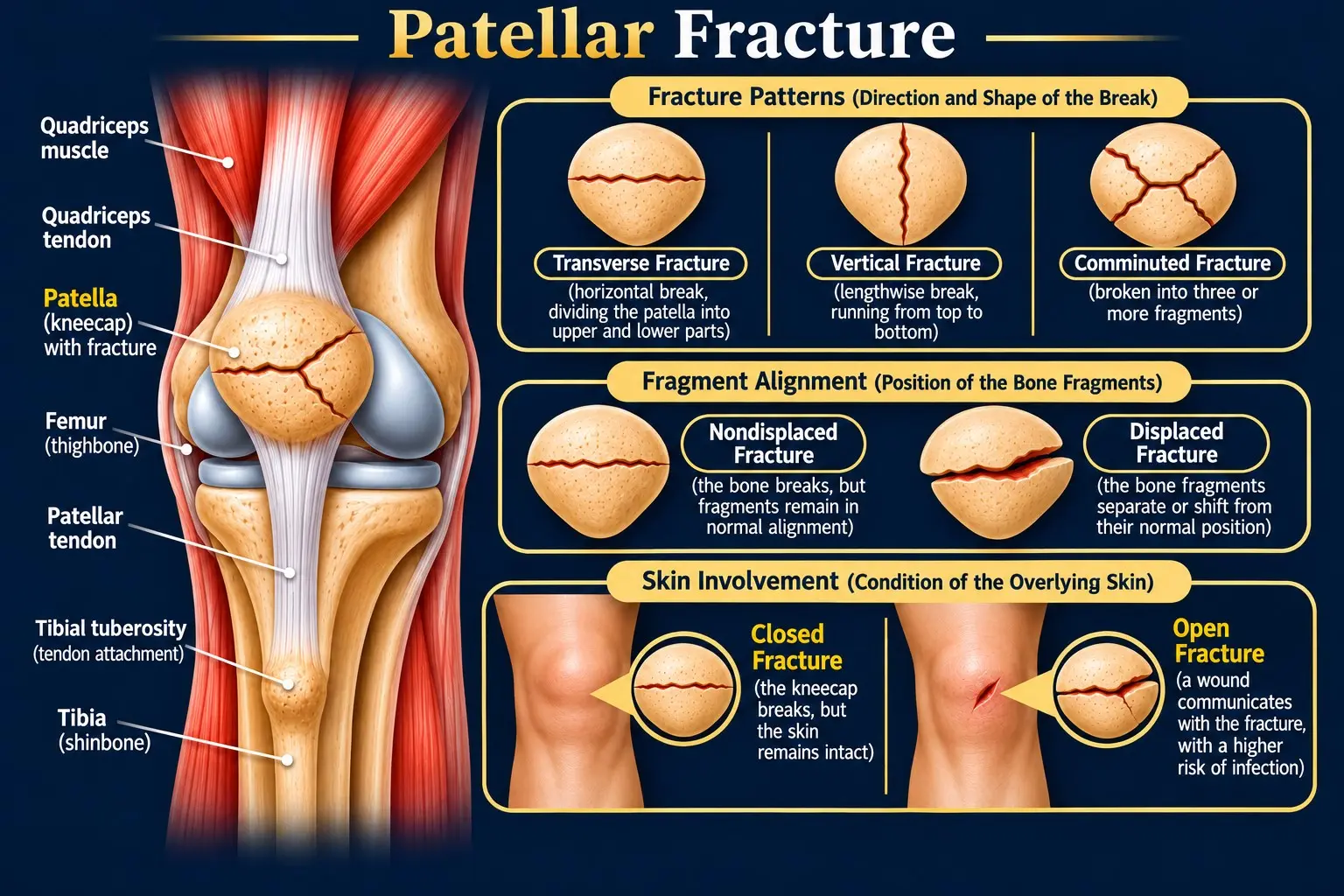
A patellar fracture most often occurs after a direct blow to the kneecap or a forceful contraction of the quadriceps muscle. The injury happens when the force exceeds the patella’s ability to withstand it.
Common causes of a patellar fracture include:
Direct trauma occurs when an external force strikes the kneecap and breaks the bone. Indirect injury occurs when a strong quadriceps contraction creates excessive tension across the patella, causing it to fracture without a direct blow.
Bone weakness, such as that associated with osteoporosis, may increase the risk of a fracture after relatively minor trauma. Most patellar fractures, though, result from significant impact or forceful muscle contraction.
Doctors classify patellar fractures by the direction of the break, the alignment of the bone fragments, and whether the skin remains intact.
These classifications describe different features of the injury and can overlap. For example, a transverse fracture may also be displaced, while a comminuted fracture may be either open or closed.
A patellar fracture can cause pain, swelling, bruising, and difficulty straightening the knee or bearing weight. Symptoms often begin immediately after a fall, direct blow, or forceful quadriceps contraction. Their severity depends on the fracture pattern, bone fragment displacement, and damage to surrounding tissues.
Symptoms vary considerably from person to person. A stable fracture may cause localized pain and swelling, while a displaced or comminuted fracture can severely limit knee movement. Associated cartilage, tendon, or soft-tissue damage may cause additional problems.
If you suspect a patellar fracture, seek prompt medical assessment, even if you can still walk. Avoid forcing the knee to bend or straighten until a healthcare professional has assessed the injury.
Doctors diagnose a patellar fracture by assessing the injury, examining knee function, and using imaging tests to identify the fracture pattern. These steps help confirm the break, determine whether bone fragments have shifted, and assess damage to the structures that straighten the knee.
The doctor asks how the injury occurred and which symptoms developed. This information helps identify the likely cause and assess the injury's severity.
Questions may include:
A direct impact or forceful quadriceps contraction may raise suspicion of a patellar fracture.
The doctor examines the knee for tenderness, swelling, bruising, deformity, and skin wounds. The examination also helps assess the extensor mechanism and identify associated injuries.
The examination may include:
An inability to straighten the knee actively may indicate damage to the extensor mechanism. Pain and swelling can also limit movement, so this finding alone does not confirm the extent of the injury. The doctor avoids forceful testing that could worsen the fracture.
X-rays are the primary imaging test for most suspected patellar fractures. Doctors usually obtain front and side views, sometimes adding other views when needed.
X-rays help determine:
These findings help the orthopedic specialist determine whether the fracture may heal with immobilization or requires surgery.
A CT scan provides detailed cross-sectional images of the kneecap. Doctors may recommend it when X-rays do not fully show a complex fracture or when several bone fragments are involved.
CT imaging helps clarify the fracture pattern, assess joint-surface involvement, and plan surgery when necessary. It is not routinely required for every patellar fracture.
Doctors may consider an MRI when they suspect associated cartilage damage, tendon injuries, ligament injuries, or other internal knee problems that X-rays cannot adequately show.
MRI is not usually the first imaging test for a straightforward patellar fracture. Ultrasound may occasionally help assess suspected tendon injuries, depending on the clinical findings.
The doctor selects additional imaging based on the physical examination and initial X-ray results. Not every patient needs a CT scan, MRI, or ultrasound.
Treatment for a patellar fracture depends on the fracture pattern, bone fragment displacement, and whether the knee’s extensor mechanism remains intact. The main goals are to restore the kneecap’s alignment, allow the bone to heal, preserve knee extension, and regain movement and strength.
Doctors generally choose between nonsurgical treatment and surgery based on the specific injury.
Nonsurgical treatment may be appropriate for stable, nondisplaced fractures when the bone fragments remain aligned and the knee’s extensor mechanism functions properly.
Follow-up examinations and X-rays help confirm that the fracture remains stable and is healing properly.
Surgery may be recommended for significantly displaced fractures, certain comminuted fractures, open fractures, or injuries that disrupt the extensor mechanism and prevent active knee extension.
Common surgical approaches include:
After surgery, rehabilitation helps restore knee movement and strength while protecting the repair. The recovery plan depends on the fracture pattern, surgical technique, and progress with healing.
The most appropriate treatment varies from person to person. An orthopedic specialist assesses the fracture and knee function before recommending a treatment plan.
A patellar fracture may require surgery when the bone fragments separate significantly or the knee’s extensor mechanism is disrupted. The surgeon also considers damage to the joint surface, associated injuries, and whether the fracture can heal in a stable position without an operation.
An orthopedic surgeon may recommend surgery in the following situations:
Nonsurgical treatment may be appropriate when the fracture is stable or nondisplaced, the joint surface remains well aligned, and the patient can actively straighten the knee.
These fractures are commonly managed with a cast, splint, or locked knee brace. Follow-up X-rays help confirm that the fragments remain aligned, while guided physiotherapy restores movement and strength when appropriate.
The decision to operate depends on the fracture’s characteristics and clinical assessment. Measurements of bone displacement can help guide treatment, but they are not absolute rules. Many stable patellar fractures heal without surgery.
Recovery from a patellar fracture depends on the fracture pattern, treatment method, bone healing, and progress with rehabilitation. Many patellar fractures need approximately 6–12 weeks for initial bone healing. Regaining full knee movement, strength, and function may take 3–6 months or longer.
These timeframes are general estimates. Not every fracture follows the same recovery pattern, and progress depends on clinical assessment and follow-up imaging.
Yes. The fracture pattern and treatment approach influence how quickly patients regain knee function.
The timing of each rehabilitation stage depends on fracture stability, follow-up imaging, and the treatment method. Your orthopedic specialist will determine when it is safe to increase knee movement, weight-bearing, and strengthening exercises.
Before returning to running, jumping, or contact sports, you generally need adequate bone healing, comfortable knee movement, sufficient quadriceps strength, and good functional control. Your treating clinician should confirm when these activities are safe.
Follow your rehabilitation plan and attend scheduled appointments. Returning to strenuous activities too early can place unnecessary stress on the healing knee.
Complications after a patellar fracture can develop from the initial injury, the healing process, or surgical treatment. Their likelihood depends on fracture severity, associated damage, treatment, and rehabilitation. Although complications are possible, many patients recover well with appropriate care.
Potential complications include:
Follow-up appointments help your clinician monitor bone healing, knee movement, strength, and possible complications. Seek medical advice if you develop worsening pain, increasing swelling, wound redness, drainage, or fever. Sudden shortness of breath or chest pain requires emergency medical attention.
Caring for your knee during a patellar fracture recovery involves protecting the fracture, managing swelling, and gradually restoring movement under medical supervision. Follow your treating clinician’s instructions because care requirements vary depending on the fracture severity and treatment method.
Contact your healthcare provider if pain or swelling worsens, your brace becomes excessively tight, or you notice signs of infection. Seek emergency medical attention for sudden shortness of breath or chest pain.
A fractured kneecap can range from a stable break to a severe injury involving displaced bone fragments or damage to the knee’s extensor mechanism. Suspected patellar fractures need prompt medical assessment to determine the severity and appropriate treatment.
Yes, some patellar fractures heal without surgery. Nonsurgical treatment may be suitable when the fracture is stable, the bone fragments remain well aligned, and the knee’s extensor mechanism functions properly. Follow-up examinations and X-rays help monitor healing.
Seek urgent medical assessment after a knee injury involving severe pain, significant swelling, visible deformity, an open wound, or difficulty straightening the knee. An open patellar fracture requires emergency medical care. Any wound near a suspected fracture needs urgent assessment, even if the bone is not visible, because the wound may communicate with the fracture.
Some people can walk with a stable patellar fracture, while others cannot bear weight safely. Being able to walk does not rule out a fracture or indicate its severity. Do not rely on your ability to walk or straighten the knee to rule out a fracture. Seek prompt medical assessment after a significant knee injury.