
Specialties › Knee & Leg › MPFL Injury

An MPFL injury affects the medial patellofemoral ligament, a strong ligament on the inner side of the knee that helps keep the kneecap stable. The injury often occurs when the kneecap moves outward during a dislocation, sudden twist, awkward landing, or direct trauma.
This article explains everything you need to know about MPFL injuries. You will learn what causes them, the different types, common symptoms, how doctors diagnose the injury, and the available treatment options. We also cover MPFL repair and reconstruction, recovery timelines, when surgery may be considered, and answers to common patient questions.
Book a consultation and receive expert orthopedic guidance.
An MPFL injury occurs when the medial patellofemoral ligament is stretched or torn, often after the kneecap moves outward. The MPFL is a strong ligament on the inner side of the knee that connects the patella, or kneecap, to the femur, or thighbone.
The ligament helps keep the patella centered within the trochlear groove, a shallow groove at the end of the femur. It provides the main soft-tissue restraint against excessive outward movement of the kneecap, especially during the early part of knee bending.
An injury can range from a mild stretch to a partial or complete tear. The severity can affect knee stability and may contribute to recurrent patellar instability when the kneecap repeatedly shifts or dislocates.
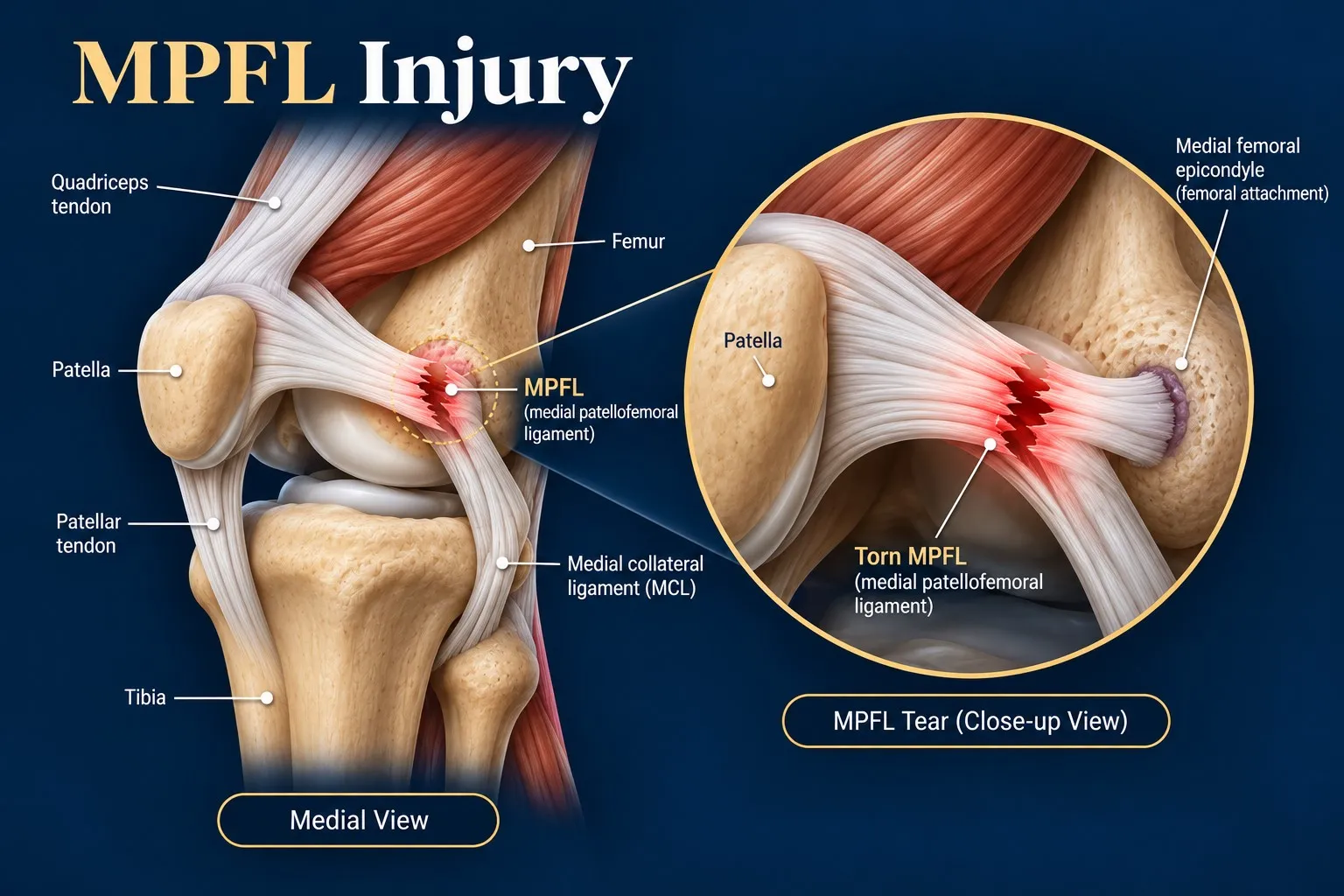
An MPFL injury most often occurs when the kneecap is forced laterally during trauma or a sudden change in movement. This can stretch, partially tear, or completely rupture the ligament.
The causes and risk factors often overlap. A sudden movement can trigger the injury, while certain anatomical or physical factors can make the kneecap less stable.
The main causes and risk factors of an MPFL injury include:
An MPFL injury can result from a single traumatic event or occur in a knee that already has factors that increase patellar instability. Identifying these factors can help guide treatment and reduce the risk of recurrent instability.
MPFL injuries can range from a mild ligament strain to a partial or complete tear. Doctors may describe the severity according to the extent of ligament damage.
Doctors may also describe an MPFL injury by its location. The tear can occur near the patella, through the middle of the ligament, or near its femoral attachment. An avulsion injury can also occur when the ligament pulls away from its attachment and takes a small piece of bone with it.
Symptoms of an MPFL injury vary according to the severity of ligament damage and the degree of patellar instability. Pain, swelling, and a feeling that the kneecap has shifted are common after an injury.
The common symptoms of an MPFL injury include:
A mild MPFL injury may cause pain, tenderness, and minimal swelling without major instability. A partial tear can cause moderate pain, swelling, and temporary feelings of kneecap instability. A complete tear may cause severe pain, significant swelling, marked instability, or patellar dislocation or recurrent subluxation.
Symptoms may become more pronounced when an MPFL injury occurs with cartilage, bone, meniscus, or other knee damage. Persistent feelings of the kneecap slipping or giving way should be evaluated by an orthopedic professional.
Doctors diagnose an MPFL injury by combining the injury history with a physical examination and appropriate imaging. The diagnostic process evaluates ligament damage, patellar stability, and any associated injuries to the bone, cartilage, or other knee structures.
The doctor asks how the injury occurred and whether the kneecap dislocated or partially shifted. They may also ask about pain, swelling, popping sensations, previous patellar dislocations, and earlier knee injuries.
The doctor checks for swelling, bruising, tenderness along the inner side of the kneecap, knee alignment, range of motion, and the ability to bear weight.
The doctor gently moves the kneecap to assess excessive outward movement. They may perform a patellar apprehension test, which can reproduce the feeling that the kneecap may slip out of position.
The doctor observes how the kneecap moves as the knee bends and straightens. Abnormal tracking or excessive outward movement may indicate patellar instability.
The examination may also check for cartilage damage, osteochondral injuries, meniscus problems, or other ligament injuries. These findings can affect treatment decisions.
X-rays can identify fractures, bone fragments, abnormal patellar alignment, and anatomical features such as a high-riding kneecap or an unusually shallow trochlear groove. X-rays do not directly show the MPFL.
An MRI provides detailed images of the MPFL and surrounding soft tissues. It can show whether the ligament is stretched, partially torn, or completely torn. MRI can also identify cartilage damage, bone bruising, osteochondral injuries, meniscus tears, and other ligament injuries.
A CT scan may be used when detailed bone anatomy or measurements are needed, particularly during surgical planning. It can help evaluate patellar alignment and the shape of the trochlear groove.
The diagnosis combines the clinical examination and imaging findings to determine the severity of MPFL damage, the degree of patellar instability, and whether associated injuries are present.
Treatment for an MPFL injury depends on the injury grade, knee stability, and associated injuries. Doctors also consider whether the kneecap has dislocated, whether instability continues, and the patient’s activity demands.
Most first-time or mild MPFL injuries can initially receive conservative treatment, particularly when there is no major bone or cartilage damage.
Surgery may be considered when non-surgical treatment does not restore stable kneecap function or when significant structural damage is present. Recurrent patellar dislocation is an important factor when considering surgery.
MPFL repair reattaches a recently torn ligament to its original attachment. It may be suitable for selected acute injuries when the ligament tissue remains suitable for repair.
MPFL reconstruction replaces the damaged ligament with a tendon graft. The graft can come from the patient's own tissue or a donor.
Reconstruction may be considered for:
A patellar dislocation can cause an osteochondral fracture or cartilage injury. Surgery may be needed to remove or fix loose bone and cartilage fragments or treat significant cartilage damage.
Some patients have anatomical factors that contribute to recurrent instability. Selected patients may need procedures such as a tibial tubercle osteotomy or other corrective procedures alongside MPFL reconstruction.
Rehabilitation remains an important part of treatment. The program progresses from controlling pain and swelling to restoring motion, strength, balance, functional movement, and eventually sport-specific activity when appropriate.
MPFL recovery time varies according to the injury grade and whether other knee structures are involved. Recovery also depends on whether treatment is non-surgical or surgical, the presence of recurrent patellar instability, and progress during rehabilitation.
| Injury or Treatment | Typical Recovery Time | General Milestones |
|---|---|---|
| Mild MPFL sprain | 2–6 weeks | Pain and swelling improve, followed by restored motion and gradual strengthening. |
| Partial MPFL tear | 6–12 weeks | Bracing and rehabilitation help restore strength, balance, and patellar stability. |
| First-time patellar dislocation, non-surgical | 6–12 weeks | Motion and strength gradually return before resuming normal activities. |
| MPFL repair | 4–6 months | Progressive strengthening and functional training precede return to sports. |
| MPFL reconstruction | 6–9 months | Running, agility, and sport-specific training progress after adequate strength and stability return. |
| Complex reconstruction or associated injuries | 9–12 months or longer | Additional bone, cartilage, or alignment procedures may extend rehabilitation. |
A mild isolated injury may recover relatively quickly, while complex injuries requiring surgery can take many months. The treating orthopedic professional and physical therapist should determine when it is safe to progress through each stage.
You may be able to walk with an MPFL injury, especially when the damage is mild. Pain, swelling, or instability can make walking difficult after a more severe injury. Being able to bear weight does not rule out significant MPFL damage or patellar instability.
Yes. An MPFL injury can occur with patellar subluxation, where the kneecap partially shifts outward without completely dislocating. Sudden twisting, pivoting, or other movements can also stress or tear the ligament without a complete kneecap dislocation.
Yes. A stretched or damaged MPFL can reduce the ligament's ability to stabilize the kneecap. Persistent MPFL insufficiency may contribute to recurrent patellar instability and repeated dislocations, particularly when other anatomical risk factors are also present.
Brace duration varies according to injury severity, treatment approach, and rehabilitation progress. A patellar-stabilizing brace may support the kneecap during healing, while the treating orthopedic professional determines when it is appropriate to reduce or stop brace use.
The provided source does not specify a fixed time for returning to driving after MPFL surgery. Driving should depend on factors such as the operated leg, mobility, ability to control the vehicle safely, pain, medication use, and medical clearance.
Exercise can be part of MPFL rehabilitation, but activity should progress gradually. Rehabilitation typically begins with restoring motion and controlling symptoms before progressing to strengthening, balance, functional movement, and sport-specific activities when appropriate.
Recurrent instability can occur after an MPFL injury, particularly when the ligament remains insufficient or anatomical factors continue to affect patellar stability. Appropriate rehabilitation and treatment of relevant structural problems can help address factors associated with recurrent instability.